DORSAL MUSCLE TIGHTNESS. Located dorsally between the transverse and spinous processes. According to the direction of the muscle fibers, it can be divided into two muscle groups.
Lateral muscle group. More laterally located muscles take their origin from the spinous processes and, going cranioventrally, are thrown over several segments and end on the transverse processes. If the muscle, having a cranioventral direction, begins on the transverse process, then in this case it ends on the transverse process of another vertebra.
The lateral group includes the longest muscle - m. Lon-gissimus, which has five parts: the longest muscles of the chest and lower back, neck, head and atlas (Fig. 100, 101).
1 and 2. The longest muscle of the chest and lower back - ni. longissimus thoracis et lumborum is the most potent part. On the lower back, it occupies a triangular space between the spinous and transverse processes. It is attached caudally along the anterior edge of the wing, ilium and spinous processes of the sacral, lumbar and last four to five thoracic vertebrae. Separate muscle bundles bifurcate and end at the transverse processes of the lumbar and thoracic vertebrae and at the vertebral ends of the ribs. The last muscle bundle comes from the spinous process and ends at the transverse process of the seventh cervical vertebra. Outside (especially in a horse), the muscle is covered with a powerful lustrous tendon, to which the gluteus medius muscle is attached in the caudal part.

Rice. 100. Deep muscles of the spinal column of a cow
3. The longest muscle of the neck - m. longissimus cervicis is located in the corner of the spine between the cervical and thoracic parts. It starts from the transverse processes of the first six to eight thoracic vertebrae and the transverse spinous fascia, emerges from under the longest muscle of the chest, and is fixed on the transverse processes of the last four cervical vertebrae. It is covered by the cervical part of the dentate ventral muscle.
4 and 5. The longest muscle of the head and atlas - m. longissimus capitis et atlantis. A narrow ribbon begins on the transverse processes of the first two or three thoracic vertebrae, emerges from under the longest muscle of the neck, goes to the head, simultaneously fixing with separate bundles on the articular processes of typical cervical vertebrae, and ends with two tendon branches along the edge of the wing of the atlas and on the mastoid process of the temporal bones. In pigs, it ends at the wing of the Atlantean and the occipital bone. In dogs, it has two parts - the longest muscle of the atlas goes from the transverse processes of three to five cervical vertebrae to the edge of the wing of the atlas, and the longest muscle of the head is well expressed, goes from the transverse processes of the 1st to 4th thoracic vertebrae and ends on the mastoid process of the temporal bone.

Rice. 101. Diagram of the dorsal muscles of the spinal column
Iliac-costal muscle - m. iliocostal lies below the longest muscle of the chest and lower back, has three parts: the lumbar, thoracic and cervical. The lumbar part is expressed in dogs and ruminants, begins on the mice and the transverse processes of the first lumbar vertebrae and ends on the last rib. The thoracic part with separate muscle bundles is fixed on the cranial edges of the upper sections of the ribs, is directed cranioventrally and ends with shiny thin tendons, throwing over 3-4 ribs, on the caudal edges of the anterior ribs and the transverse process of the 7th cervical vertebra. The cervical part is located below the dentate ventral muscle. Fixing in separate bundles on the transverse processes of the cervical vertebrae, it goes, throwing over the segment, up to the 3-4th vertebra. In pigs it comes to Atlantis,
Plaster muscle - m. splenitis is located in a wide triangular layer in the neck, filling the space in herbivores between the vertebrae and the columnar part of the nuchal ligament. It is covered in the caudal part by the dentate ventral and rhomboid muscles. It originates at the withers on the third, fourth and fifth spinous processes and transverse spinous fascia. Dorsally along the course, it is fixed with tendon bundles on the columnar part of the nuchal ligament and, expanding in a fan-like manner, is directed cranioventrally, ends with separate teeth on the transverse processes of the cervical vertebrae, the wing of the atlas, the crest of the occipital bone and the mastoid process of the temporal bone (in ruminants and dogs - only on the occipital bones).
In ruminants, along the way, it is fixed on the 1st and 2nd cervical vertebrae.
In horses - on the 1st, 3rd - 5th neck, in the case of the neck - on the first. Both have no cervical attachment. All muscles of the lateral group are innervated by the dorsal branches of the cervical spinal nerves.
Medial muscle group. Located under the lateral muscle group of the dorsal cord of the spinal column. The direction of the muscle fibers is craniodorsal. The muscle bundles of this muscle group go from the transverse processes to the spinous processes or along the spinous processes.
Spinous muscle of the chest and neck - m. spinalis thoracis et cervicis. Developed more at the withers. On the lower back, it merges at the base of the spinous processes with the longest muscle of the lower back and increases its mass where the longest muscle ends. Anchored on the spinous processes of the withers, from the bases of the transverse processes to their tops, it extends onto the spinous processes of the cervical vertebrae. Each bundle of it is thrown over several segments. This is one of the deepest muscles.
On the neck of herbivores, it lies on the lamellar part of the nuchal ligament.
In pigs, it goes up to the 6th (5th) cervical vertebra.
In dogs it ends in the last two cervical vertebrae.
Semispinal muscle of the head - m. semispinalis capitis. Located in the neck, has the shape of an elongated triangle, well expressed. Covered by a plaster muscle. At its base there is a tape of the longest muscle of the head. It begins in herbivores on the first five to eight transverse processes of the thoracic and last cervical vertebrae and is fixed on the occipital bone on the side of the nuchal ligament.
In pigs and dogs, it is divided into two parts - dorsal and ventral.
Divided muscles - mm. niultifidi. These are the deepest muscles on the dorsal side of the spinal column. In short bundles, they go in the craniodorsal direction, throwing over several segments, from the transverse to the spinous processes, directly covering the arches of the vertebrae. In the caudal part of the spine, they have longer bundles, anchored craniodorsally at the apexes of the spinous processes. In the cranial part, they are fixed at the base of the spinous processes.
In herbivores (especially ruminants), they are well developed on the neck, going from the articular processes to the spinous processes of the typical cervical vertebrae.
In dogs, strongly developed up to the crest of the axial vertebra.
To better understand the topography of the dorsal muscles of the spinal column in the neck, distribute them in layers. There are five layers and two muscles in each layer. The first two layers are the cervical parts of the muscles that hold the shoulder girdle: 1) the trapezius muscle, along its lower edge lies the brachiocephalic muscle; 2) the dentate ventral (cervical part), along its upper contour, the rhomboid muscle lies above. The third layer is the plaster muscle, along its ventral edge in the lower third of the neck is the longest neck muscle. The fourth layer is the longest muscle of the head and above is the semispinal muscle of the head. The fifth layer is the multifidus muscles and the spinous muscle of the chest and neck.
To facilitate the study of the muscles of the spinal column, short muscles are separately highlighted on it. They are located more on the most mobile parts of the spinal column - the neck and tail, and much less on the lower back. These are short interspinous and intertransverse muscles, as well as short head muscles and short tail muscles.
Intertransverse muscles - mm. intertransversarii, according to their attachment, are divided into dorsal ones, running cranioventrally from the articular processes of the neck vertebrae to the transverse ones; middle, fixed between the transverse processes of the cervical vertebrae, and ventral, fixed between the transverse processes of the lumbar and costal processes of the cervical vertebrae,
In pigs, the lateral part of the intertransverse muscle forms a prominent solid long strand that runs along the neck to the atlas.
Interspinous muscle - m. interspinal. Located between the spinous processes, well developed in dogs and pigs in the cervical, thoracic and lumbar regions. In herbivores, it has evolved into static muscles (ligaments).
Short muscles of the head. Located in the region of the first two joints of the neck - atlantooccipital and atlantoaxial.
Short muscles of the head acting on the atlantooccipital and atlantoaxial joints.

The short muscles of the head provide movement of the head near the atlas (right, left, up and down) and the movement of the head with the atlas near the axial vertebra (bottom up, right and left), rotation occurs along the sagittal axis. In the area of these joints, there are two paired oblique and four paired rectus head muscles.
On the dorsal side of the atlas, two oblique head muscles go, forming a rhombus: the caudal oblique muscle of the head - m. obliquus capitis caudalis (from the crest of the axial vertebra, both symmetrical parts of this muscle are directed by wide fleshy plates and are fixed along the entire edge of the wing of the atlas; the direction of the fibers is craniolateral) and the cranial oblique muscle of the head - m. obliquus capitis cranialis (a narrow abdomen goes from the cranial edge of the wing of the atlas to the bases of the jugular processes of the occipital bone, in horses - to the occipital ridge, in dogs - to the mastoid process).
The dorsal rectus muscle of the head is large - m. rectus capitis dorsalis major goes from the crest of the axis to the occipital bone, fused with the semispinal muscle of the head. It can go in two layers.
The dorsal rectus muscle of the head is small - m. rectus capitis dorsalis minor, covered by the dorsal large head muscle, lies directly on the capsule of the atlantooccipital joint. Goes from the dorsal arch of the atlas to the occipital bone.
Lateral rectus muscle of the head - m. rectus capitis lateralis is located below the atlas from its ventral arch and ends on the jugular processes.
Ventral rectus muscle of the head - m. rectus capitis ventralis lies below the atlantooccipital joint, goes from the ventral arch of the atlas to the body of the occipital bone.
Both ventrally located muscles no longer belong to the dorsal muscle cord and are innervated by the ventral branches of the cervical spinal nerves. Described in this section for the convenience of studying the material and a complete understanding of the muscles acting on the first two joints of the neck.
Tail muscles. On the tail, the muscles are located on the dorsal part (long and short tail lifters), laterally (intertransverse tail muscle) and on the ventral part (long and short tail lifters and tail muscle). The latter already belong to the ventral muscles of the spinal column and are described here only for the convenience of memorization.
Short tail elevator (medial dorsal sacro-caudal muscle - m. Sacrocaudalis dorsalis medialis). It starts from the spinous processes of the last sacral and first caudal vertebrae, throwing over the segment, and ends at the rudiments and articular processes of the caudal vertebrae.
Long tail elevator (lateral dorsal sac-tail muscle - m. Sacrocaudalis dorsalis lateralis). Located lateral to the previous one. It goes from the intermediate crest of the sacral bone and from the dorsal surface of the transverse processes of the caudal vertebrae and ends in thin tendons, throwing over several segments, on the rudiments and articular processes of the caudal vertebrae.
In pigs, the tendons of the tail lifters are spirally twisted, giving the tail a corkscrew appearance.
In dogs and pigs, these muscles start from the last lumbar vertebrae.
Dorsal intertransverse muscles of the parasite - mm. intertrausversarii dorsales caudae are fixed between the rudiments of the transverse processes and disappear with the latter. Well developed in cattle.
The long tail tail (lateral ventral sacro-tail muscle - m. Sacrocaudalis ventralis lateralis) lies laterally, and the short tail tail (medial ventral sacro-tail muscle - m. Sacrocaudalis ventralis medialis) is medial. Both originate, respectively, on the sacrum and end on the transverse processes and the bodies of the caudal vertebrae. In pigs, they are poorly expressed.
In pigs and dogs, it starts from the last lumbar vertebrae.
Caudal muscle - m. coccygeus - ribbon-like, originates on the medial surface of the ischium and ends on the transverse processes of the first caudal vertebrae. Along the way, it is adjacent to the lateral wall of the rectum. While contracting, it presses the tail to the anus, not simultaneously - pulls the tail to the side.
VENTRAL MUSCULAR TENSION. These muscles are located on the ventral surface of the vertebral bodies and transverse processes, mainly in the neck, loin and tail.
Long neck muscle - m. longus colli has a chest and neck. On the thoracic part, each muscle bundle (right and left) begins on the body of the 5-6th thoracic vertebra, goes craniolaterally to the transverse processes of the 6-7th cervical vertebra, forming sharp angles directed caudally. The cervical part of this muscle also begins with separate teeth from the ventral surface of the transverse processes and bodies of the last five cervical vertebrae, which are directed craniomedially to the ventral crests of the vertebrae to the ventral arch of the atlas, throwing over several segments. The right and left muscles form sharp angles directed cranially so that at the junction of the cervical spine in the pectoral region, these muscles form a rhombus. It's easy to find this muscle here.
Long muscle of the head - m. longus capitis runs on the sides of the long neck muscle, forming a ridge. It begins on the ventral surfaces of the transverse processes of the 2nd-6th cervical vertebrae and ends on the body of the occipital bone. The two ventral rectus muscles of the head are described in the short head muscles group.
On the ventral surface of the lumbar region is the square psoas muscle - ni. quadratus lumborum, formed by interconnected muscle bundles. It goes from the medial surface of the vertebral ends of the last two ribs, fixing on the ventral surface of the transverse processes of the lumbar vertebrae, and ends on the ventral surface of the wings of the sacral bone.
The psoas quadratis muscle is covered by the greater and medially lying psoas minor muscles, which are described in the Muscles and Fascia of the Pelvic Extremity section. The ventral muscles of the tail are described above (see “Muscles of the tail”).
The entire musculature of the dorsal cord is innervated by the dorsal, and the ventral cord is innervated by the ventral branches of the spinal nerves.
The ventral region of the neck is represented by the larynx, trachea, esophagus, thyroid gland, as well as the muscles and fascia that surround them. Since the location of these organs is not the same in different regions of the neck, the surgical manipulations in the ventral region of the neck are very diverse.
In the ventral region, six layers are distinguished.
- The skin is thin and mobile with a small layer of subcutaneous tissue, in places it is tightly connected to the cutaneous muscle of the neck.
- Superficial fascia, cutaneous muscle of the neck with longitudinally directed fibers.
- The brachiocephalic muscle (in the lateral region of the neck) and the sterno-jaw muscle (in the lower-lateral region of the neck). The gap between them is called jugular groove.
- The brachiohyoid muscle is connected in the posterior and middle part almost inseparably with the brachiocephalic muscle. In the front third of the neck and in the parotid region, it closely fuses with the sternohyoid muscle, but is separated from the brachiocephalic muscle.
- The sternohyoid and sterno-thyroid muscles are located in the lower part of the neck. The visceral fascia covers the sides of the neck organs. These muscles are enclosed in the parietal layer of the visceral fascia, which surrounds the larynx and pharynx cranially, and is caudally attached to the ribs and hilt of the sternum at the entrance to the chest cavity.
- Actually the organs of the neck, each of which is surrounded by its own fascia. They are connected by jumpers both between themselves and with the parietal leaf of the visceral fascia, which acts as a support for all fascia and a common cover for the organs of the neck.
Trachea located ventrally from the long muscle of the neck. From the larynx to the entrance to the chest cavity, its cervical part extends. The basis of the trachea is made up of open cartilaginous rings. From the dorsal side, the ends of the rings are connected by the transverse connective tissue ligament. The mucous membrane is loosely connected with the lower and lateral walls of the trachea. The fascia of the trachea, esophagus and the neurovascular bundle are connected. The cervical part of the trachea is quite mobile, especially in the lateral directions.
The blood supply to the trachea is carried out by short tracheal branches of the common carotid artery, which form longitudinal arches. Segmental inter-annular vessels are connected in the midline with the branches of the same name on the other side.
The branches of the vagus and sympathetic nerves innervate the trachea.
Esophagus begins with the pharyngeal opening, located dorsally from the trachea. At the level of IV, the cervical vertebra deviates to the left and, before entering the chest cavity, follows the left upper-lateral edge of the trachea. At the level of the VII cervical vertebra, the esophagus is again located dorsally from the trachea and thus penetrates into the chest cavity. Outside, the cervical part of the esophagus is covered with adventitia (connective tissue sheath), firmly connected with the muscle layer. The mucous membrane is dense, extensible, connected to the muscle layer by loose fiber; at rest, it is usually collected in longitudinal folds. The cervical part of the esophagus is covered with its own fascia. Both the thickness of the wall and the diameter of the lumen of the esophagus in different departments are not the same. Adjacent to the esophagus is the left common carotid artery, the left tracheal lymphatic duct, the vagosympathetic trunk and the recurrent nerve.
The cervical esophagus is supplied with blood from the short branches of the common carotid artery and the cranial thyroid artery. The branches of the vagus, sympathetic and glossopharyngeal nerves innervate the esophagus.
Birds have a thickening of the esophagus - goiter... In chickens and pigeons, the goiter is represented by a bag-like subcutaneous protrusion: single (in chickens) and paired (in pigeons). In geese and ducks, the crop looks like a longitudinal expansion.
The jugular groove is formed by the brachiocephalic (upper wall), sterno-jaw (lower wall) and shoulder-hyoid (bottom of the groove) muscles.
The jugular vein is located in the jugular groove. At the level of the II cervical vertebra, it is formed by the fusion of the external and internal jaw veins. Before entering the chest cavity, both jugular veins merge into the bibular trunk, which flows into the anterior vena cava. The diameter of the external jugular vein, when filled, can reach 3 cm. Valves are located on its inner surface. The first pair of valves is located at a distance of 15 cm from the confluence of the external and internal jaw veins; this is level IV of the cervical vertebra. Medicines are injected into the area of the vein, free of valves, and blood is taken from it. The jugular vein is covered by the fascia of both the thin own and passing from the sterno-brachiocephalic muscle. In the sheets of its own fascia, the cutaneous nerve of the neck runs along the dorsal surface of the vessel, the upper-inner edge of the groove.
Blood supply to the ventral neck carries the common carotid artery (through numerous branches) and the ascending cervical artery.
The neck skin is innervated by the ventral branches of the cervical nerves. The cutaneous muscles of the neck innervate the cutaneous nerve of the neck, formed by the cervical branch of the facial nerve and the ventral branch of the 2nd cervical nerve. The sterno-jaw muscle is innervated by the accessory nerve. The sternohyoid, sterno-thyroid and brachiohyoid muscles are innervated by the branches of the ventral cervical nerves.
If you find an error, please select a piece of text and press Ctrl + Enter.
MUSCLES OF THE NECK AND SHOULDER Girdle (mm.cevicis et cinguli membri thoracici)
According to the places of attachment, location and function, these muscles can be divided into two groups: muscles that are anchored along the dorsal contour of the trunk (dorsal attachment), and muscles that are attached along the ventral contour (ventral attachment).
These muscles are mostly lamellar and are located on the neck, scapula, and chest. They have one attachment point on the trunk, and another on the scapula and humerus. The muscles of the shoulder girdle provide the forward or backward movement of the pectoral limbs, and also assist in flexion, extension, and rotation of the limb at the shoulder joint.

Muscles of the trunk and shoulder girdle of a horse
Dorsal anchorage muscles.
The most superficial of them is the trapezius muscle - m. trapezius is located directly under the skin in the neck and withers. It is thin, lamellar and has the shape of a versatile triangle, which with its base is directed to the dorsal midline, and its apex is directed to the spine of the scapula. In the muscle, the cervical and chest parts are distinguished. Both begin with a lamellar tendon along the nuchal and supraspinous ligaments.
The cervical part - pars cervicalis - with its wide tendon edge originates from the cord of the nuchal ligament in the horse at the level of the 2nd cervical and up to the 3rd thoracic vertebra. The muscle bundles, converging ventrocaudally, end along the entire cranial edge of the scapula spine.
The pectoral part - pars thoracica - is more fleshy, shorter and narrower than the previous one. It is attached to the supraspinatus ligament in the range from the 3rd to 10th thoracic vertebrae and, narrowing, goes to the scapula, ending only on the dorsal part of the scapula spine.
The muscle strengthens the scapula on the trunk. With a unilateral contraction, it contributes to the bending of the neck and the extension of the limb forward.
It is innervated by the XI pair of cranial nerves - the dorsal branch of the accessory nerve - r. dorsalis n. accessorii.
Rhomboid muscle - m. rhomboideus lies under the trapezius and is almost entirely covered by it. It begins at the level of the 3rd cervical vertebra and extends to the 8-9th thoracic vertebra. At the level of the 3rd thoracic vertebra it is also divided into the cervical and thoracic parts, which go under the scapula, ending on the medial side of the base of the scapula and scapular cartilage.
The rhomboid muscle of the chest - t. Rhomboideus thoracicus - is located under the initial part of the semispinal muscle of the head and partially above the cranial dorsal dentate muscle. Its muscle bundles, having a vertical direction in relation to the scapula, originate from the supraspinatus ligament in the range from the 3rd cervical to the 8-9th thoracic vertebrae. The muscle ends on the medial surface of the scapular cartilage and, dropping partially to the dorsal edge of the scapula, covers the dorsal areas of the subscapularis and ventral dentate muscles.
The rhomboid muscle of the neck - t. Rhomboideus cervicis - is located above the patch muscle. It starts from the cord of the nuchal ligament along the length from the 2-3rd cervical to 2-3rd thoracic vertebra and ends on the medial surface of the cranial angle of the scapular cartilage. In their course, the muscle bundles take a caudoventral direction and an almost horizontal position.
Both muscles are fixed on the scapula, during movement, their parts pull it forward or backward.
It is innervated by the dorsal branches of the cervical nerves and from the cranial thoracic nerves of the brachial plexus - rr. dorsales pp. cervicales et thoracici.
Latissimus dorsi muscle - m. latissimus dorsi - wide, triangular in shape, its wide base originates from the thoracolumbar fascia along the supraspinatus ligament from the 3-4th thoracic vertebra to the last lumbar vertebra (in the cranial part it is located under the rhomboid muscle). Caudodorsally, the muscle is partially covered by the subcutaneous muscle of the trunk, and dorsoventrally - by the thoracic part of the trapezius muscle. Throwing over the caudal edge of the scapular cartilage, the latissimus dorsi muscle, narrowing in the cranioventral direction, goes under the triceps muscle of the shoulder, goes under the scapula, connects with the caudoventral edge of the large round muscle and its distal tendon, and then ends at the large round roughness of the humerus.
Function - pulls the free section of the limb back, and with the supporting position of the limbs, pulls the body forward.
It is innervated from the brachial plexus - n. Thoracodorsal.
Brachiocephalic muscle - m. brachiocephalicus - wide, lamellar, fixed on the head (on the mastoid process of the temporal bone, occipital crest) and on the 2nd, 3rd and 4th cervical vertebrae, goes along the lateral surface of the neck along the ventral edge of the trapezius muscle, caudoventrally passes to the craniolateral surface of the shoulder joint and ends at the crest of the humerus. Throughout its entire length, the brachiocephalic muscle covers the plaster-like, brachio-graft, cervical part of the ventral dentate and intertransverse muscles.
The muscle is complex, it also distinguishes between the clavicular-brachialis (covers the front of the shoulder joint) and the clavicular-mastoid (attached to the mastoid process of the temporal and nuchal crest of the occipital bone; forms the dorsal border of the jugular groove) muscles.
The muscle contributes to the extension of the limb forward and unilateral flexion of the neck. With the supporting position of the limbs, its simultaneous bilateral contraction produces neck flexion, lowering and stretching of the head forward.
The complexity of the muscle is proved by its innervation: from the XI pair of cranial nerves - n. accessorius and from the brachial plexus - n. axillaris.
Muscles of the ventral anchorage.
Somewhat below and under the brachiocephalic muscle runs along the ventrolateral surface of the neck a number of muscles that do not strengthen the shoulder girdle near the body, but are fixed in the area of the scapula and sternum. These muscles are directed to the head and affect the movement of the mandible, larynx, and tongue.
Sterno-jaw muscle - m. sternomandibularis - has a ribbon-like shape, originates on the handle of the sternum under the brachiocephalic muscle. A long rounded tape is directed along the trachea, forming, together with the brachiocephalic muscle, a deep groove - the jugular groove - sulcus jugularis, in which a large external jugular vein passes. Ends on the body of the hyoid bone.
With unilateral contraction, it bends the neck, with bilateral contraction, it helps lower the neck and stretch the head, as well as opening the mouth opening.
It is innervated by the XI pair - n. accessorius.
The next three muscles - the sternohyoid, the sterno-thyroid, and the shoulder-sublingual - act on the tongue and larynx. Anchored to the hyoid bone and larynx, they stretch in the form of ribbons along the neck along the trachea, covered by the brachiocephalic and sterno-mandibular muscles, and end at the handle of the sternum and fascia. These muscles are described below in the study of the muscles of the tongue and larynx - they are mentioned here in connection with their topography.
Serrated ventral muscle - m. serratus ventralis - the main muscle on which the body is suspended between the shoulder blades, powerful, looks like a fan, spread between the medial surface of the shoulder blade, neck and chest wall. It distinguishes between two parts - the ventral serratus muscle of the neck and the ventral serratus muscle of the chest.
The ventral serratus muscle of the neck - t. Serratus ventralis cervicis - originates with the teeth from the transverse processes (3) of the 4-7th cervical vertebrae and ends on the medial surface of the cranial angle of the scapula, visible on the neck below the ventral edge of the rhomboid muscle.
With a fixed limb, the cervical parts, contracting, simultaneously raise the neck, with a one-sided contraction, they turn to the side.
Innervated from the brachial plexus nn. dorsales scapulae et n. thoracicus longus.
The ventral serratus muscle of the chest - t. Serratus ventralis thoracis - originates with muscle teeth on the lateral surface of the first nine ribs and ends on the medial surface of the caudal angle of the scapula. The teeth of the external oblique abdominal muscle are wedged between the teeth on the costal surface. From above, the muscle fuses tightly with the dentate fascia, which is highly developed in the horse.
The muscle supports the trunk between the limbs.
Innervated from the brachial plexus n. thoracicus longus.

Horse muscles on the ventral side
Pectoral muscles.
They are located under the skin on the ventral surface of the chest between the chest limbs, in the form of two layers. The pectoral muscles bring the limbs to the trunk. During movement, their cranial parts help to push the limb forward, the caudal ones - backward.
Superficial pectoral muscle - m. pectoralis superficialis - located under the skin on the ventral surface of the chest and straightened between the sternum and chest limbs. It is subdivided into the descending and transverse pectoral muscles.
The descending pectoral muscle - so the pectoralis descendens is powerful, originates from the handle of the sternum, is thrown over the shoulder joint and is fixed on the large tubercle and crest of the humerus. Contacting medially with the muscle of the same name on the opposite side, it participates in the formation of the ventral midline - linea mediana ventralis.
The transverse pectoral muscle - so pectoralis transversus - is flatter than the previous one, starts from the body of the sternum in the range from the 1st to the 6th costal cartilage and ends below the elbow joint on the fascia of the medial surface of the forearm.
Both muscles are adductors of the thoracic limb, promote the forward movement of the limb, and with the supporting position of the limbs, pull the body forward.
Deep pectoral muscle - m. pectoralis profundus (or ascending pectoral muscle - m. pectoralis ascendens). Located under the superficial pectoral muscle, it starts from the body of the sternum, abdominal fascia and 5-9 costal cartilages and ends on the large and small tubercles of the humerus, partly on the fibrous sheath of the proximal tendon of the biceps brachii; as well as on the cranial surface of the primordial muscle.
Helps superficial pectoral muscles.
Innervation: pp. pectorales craniales et n. pectoralis caudalis.
Subclavian muscle - m. subclavius. It is located cranially to the previous one, closely fuses with it, is fixed on 1-4 costal cartilages and goes around the joint and the anterior edge of the scapula. Ends on the fascia of the primordial muscle.
Innervation: pp. pectorales craniales et n. pectoralis caudalis.
This muscle group is developed only in the mobile parts of the spinal column - the cervical, lumbar, and caudal. They are antagonists of the dorsal muscles and bend the spinal column, lower the neck with the head, tail, bend the lower back. Unilateral contraction of the right and left dorsal and ventral muscles of the spinal column pulls the neck and tail to the side. The joint simultaneous action of all the dorsal muscles strains the spinal column.
Muscles located in the neck
Long muscle of the head - m. longus capitis - lies on the ventral surface of the cervical vertebral bodies, lateral to the long neck muscle. It starts from the costal processes (6-2nd) of the cervical vertebrae and ends on the muscular tubercle of the base of the skull.
Function: lowers the head and promotes lateral neck movements.
Long neck muscle - m. longus colli - divided into cervical and chest parts. Lies on the ventral surface of the bodies of the cervical and first thoracic vertebrae between the long muscles of the head. The cervical part begins on the ventral crests of 2-5 cervical vertebrae and the ventral tubercle of the atlas. It ends on the ventral surface of the bodies of 2-6 cervical vertebrae and the last tooth ends on the costal process of the 6th cervical vertebra. The thoracic part begins on the ventral crests of the 1st to 5th thoracic vertebrae and ends on the ventral surface of the bodies of the thoracic and 6-7 cervical vertebrae.
Function: bends the neck.
The rectus lateral muscle of the head - m. rectus capitis lateralis - starts from the ventral arch and pterygoid fossa of the atlas and ends at the jugular process of the occipital bone.
The rectus ventral muscle of the head - m. rectus capitis ventralis - starts from the ventral tubercle of the atlas and ends on the body of the occipital bone.
Function: lowers his head.
Muscles located in the lumbar region
Square muscle of the lower back - m. guadratus lumborum - lies on the ventral surface of the transverse costal processes of the lumbar vertebrae, covered by the psoas major muscle. It begins on the medial surface of the last 2 ribs, along the way it is attached to the transverse costal processes of the lumbar vertebrae and ends on the ventral surface of the wing of the sacral bone.
Function: participates in flexion, fixation and lateral bending of the spine.
Small psoas muscle - m. psoas minor - lies on the ventral surface of the lumbar vertebrae. It begins on the bodies of the last 3 thoracic vertebrae and the first 4-5 lumbar vertebrae. It ends at the lumbar tubercle of the ilium.
Features and Function: more developed in pigs and ruminants. Flexes the lower back, pulls it forward.
Psoas major muscle - m. psoas major - located on the ventral surface of the transverse costal processes and the bodies of the lumbar vertebrae. It begins on the medial surface of the last 2 ribs and ends on the lesser trochanter of the femur.
Features and Function: in a dog it begins only on the lumbar (3-4) vertebrae. Flexes the lower back and hip joint, participates in the forward movement of the limb.
Tail muscles
Caudal muscle - m. coccygeus - starts from the ischial spine of the pelvic bone, runs along the lateral side of the tail and attaches to the transverse costal processes of the caudal vertebrae (3-4).
Function: provides lowering and lateral tail abduction.
Medial ventral sacro-caudal muscle - m sacrocaudalis ventralis medialis - lies medially, starts from the ventral ridge of the sacral bone and ends on the hemal processes of the caudal vertebrae.
Lateral ventral sacro-caudal muscle - m. sacrocaudalis ventralis medialis - begins on the ventral surface of the sacrum and ends on the transverse costal processes and the bodies of the caudal vertebrae.
Peculiarities: in dogs and pigs, it starts from the last lumbar vertebra.
VENTRAL NECK MUSCLES
The sterno-mastoid muscle - m. Sternomastoideus begins at the handle of the sternum and ends at the mastoid process of the temporal bone.
Features and Function: the horse is absent. In a dog, it also ends in the occipital bone. He lowers his head, with one-way movement pulls his head in his direction.
Pectoralis muscle - m. sternomandibularis - covered by the cutaneous cervical muscle. It starts from the hilt of the sternum.
Features and Function: in a dog, a sheep is absent. In a horse, it ends at the caudal edge of the ramus of the lower jaw. Lowers the lower jaw. In ruminants, it ends on the facial tubercle of the maxillary bone. Lowers his head.
Sternohyoid muscle - m. sternohyoideus - lies on the ventral surface of the trachea, covered by the sternomastoid muscle. It starts from the handle of the sternum, ends on the body of the hyoid bone.
Features and Function: the dog is highly developed. Pulls the tongue back when swallowing.
Brachiohyoid muscle - m. omohyoideus - lies in the cranial part of the neck under the sternohyoid muscle, ends on the body of the hyoid bone.
Features and Function: the dog is absent. In pigs and horses, it starts from the subscapularis fascia at the distal end of the scapula. In ruminants, it starts from the deep cervical fascia. Pulls the hyoid bone back.
Sterno-thyroid muscle - m. sternothyreodeus - lies on the ventral surface of the trachea. It starts at the hilt of the sternum. It ends on the thyroid cartilage of the larynx.
Function: pulls the larynx back after swallowing.
MUSCLES OF THE LIMBS
The limbs have the following muscle groups: extensors and flexors, abductors and adductors, instep supports and pronators (rotators).
Flexors (flexors) are located inside the angle of the joint, when they contract, they reduce the articular angle, that is, they flex the joint.
Extensors (extensors) pass through the apex of the joint angle, when they contract, they increase the articular angle, while unbending the joint. The axis of movement lies horizontally in the segmental plane, and the movement of the limb occurs in the sagittal plane. On the limbs, extensors and flexors, as a rule, lie with their abdomens proximal to the joint on which they act.
Abductors (abductors) remove one limb from the other and are located on the lateral surface of the joint.
Adductors (adductors) produce the opposite effect - they bring the limbs closer and lie on the medial surface of the joint. Adductors and abductors act around a horizontal axis, which lies in the sagittal plane, and the movement of the limb in the segmental plane.
The instep supports rotate the limb outward, that is, to the lateral side.
Pronators rotate the limb in the opposite direction, while the dorsal surface of the limb is rotated medially. The action of the instep supports and pronators takes place around the longitudinal axis.
MUSCLES OF THE CHEST
Shoulder muscles
(picture 5)
In a simple, polyaxial shoulder joint, extension and flexion, abduction and adduction, and a slight rotation of the free part of the limb are possible. The abdomens of most of the muscles acting on this joint lie proximal to the joint - on the scapula.
The supraspinatus muscle - m. supraspinatus - fills the precautionary fossa of the scapula. It starts from the anterior fossa of the scapula and ends in two legs on the lateral and medial tubercles of the humerus.
Features and Function: unbends the shoulder joint.
Deltoid muscle - m. deltoideus - divided into two parts: acromial and scapular. The acromial part starts from the acromion of the scapula, the scapular - from the spine of the scapula and the extraspinatus muscle. Both parts end at the deltoid roughness of the humerus.
Features and Function: in horses and pigs, the acromial part is absent. Flexes and supines the shoulder joint.
The posterior muscle - m. infraspinatus - begins in the extraspinal fossa of the scapula and ends at the lateral tubercle of the humerus. From above it is covered by the deltoid muscle.
Features and Function: in ungulates, it performs the function of the lateral ligament of the shoulder joint. Abductor.
Small round muscle - m. teres minor - laterally covered by the deltoid muscle. It begins at the distal third of the caudal edge of the scapula and ends at the crest of the humerus under the lateral tubercle.
Features and Function: in a dog an instep support for the shoulder joint, in other animals - a flexor.
Coracoid - brachial muscle - m. coracobrachialis - located on the medial surface of the shoulder joint and humerus. It begins on the coracoid process of the scapula and ends on the medial surface of the proximal third of the humerus.
Features and Function: adductor and pronator of the shoulder joint.
Subscapularis muscle - m. subscapularis - fills the subscapular fossa, where it begins, ends at the medial tubercle of the humerus.
Features and Function: in ungulates, it functions as the medial ligament of the shoulder joint. Adductor.
Big round muscle - m. teres major - covered by the triceps muscle of the shoulder. It starts from the proximal half of the caudal edge of the scapula and ends at the circular roughness of the humerus.
Features and Function: in ungulates - flexor, in carnivores - pronator of the shoulder joint.
Figure 5 Muscles of the chest limb of a horse from the medial surface
1 - the broadest muscle of the back; 2 - large round muscle; 3 - long head of the triceps muscle; 4 - medial head of the triceps muscle;
5 - subscapularis muscle; 6 - prescapular part of the deep pectoral muscle; 7 - the shoulder of the deep pectoral muscle; 8 - karacoid shoulder muscle;
9 - biceps brachii.
Elbow muscles
(picture 1 and 5)
In a simple uniaxial elbow joint, only flexion and extension are possible, and in a dog, in addition, rotation of the forearm. The abdomens of all muscles acting on this joint are located in the shoulder region, with the exception of the pronators and instep supports, which are located distal from the joint.
Triceps muscle of the shoulder - m. triceps brachii - fills the space between the caudal edge of the scapula and the humerus. It has three heads: a long head - caput longum - starts from the caudal edge of the scapula; lateral head - caput laterale - starts from the neck of the humerus; the medial head - caput mediale - from the middle third of the humerus. All three heads end at the elbow tubercle.
Features and Function: the dog and the pig also have an additional head, which starts from the neck of the humerus, the long head flexes the shoulder joint, all heads extend the elbow joint.
Elbow muscle - m. anconeus - starts from the edges of the ulnar fossa of the humerus and ends at the ulnar tubercle. Covered by the triceps muscle of the shoulder.
Features and Function: unbends the elbow joint.
Forearm fascia tensioner - m. tensor fasciae antebrachii - located on the medial surface of the long head of the triceps brachii muscle, along its caudal edge. It begins tendonally at the caudal edge of the scapula, on the fascia of the teres major, and ends at the ulnar tuberosity and the fascia of the forearm.
Features and Function: in ungulates, this muscle acts on two joints. Extends the elbow and flexes the shoulder joint and tightens the fascia of the forearm.
Biceps muscle of the shoulder - m. biceps brachii - in the proximal part, it is covered by the brachiocephalic muscle, located on the anterior surface of the shoulder. It begins with a powerful tendon at the tubercle of the scapula and ends at the roughness of the radius.
Features and Function: in cattle and horses, a tendon cord, lacertus fibrosus, departs from the distal end of the muscle, heading to the tendon of the radial extensor of the wrist. This connects the shoulder, elbow and wrist joints. Flexes the elbow and unbends the shoulder joints.
Shoulder muscle - m. brachiialis- begins under the head of the humerus and ends at the roughness of the radius.
Features and Function: flexes the elbow joint, in carnivores it penetrates the elbow joint.
Instep support - m. supinator - located on the dorsal surface of the elbow joint, covered with the radial extensor of the wrist and the common extensor of the fingers. It starts from the lateral epicondyle of the humerus, goes obliquely and ends on the medial surface of the upper third of the radius.
Features and Function: in cattle and horses it is absent, it reaches the greatest development in ruminants. Supine forearm.
Round pronator - m. pronator teres - starts from the medial epicondyle of the humerus, ends on the medial surface of the proximal third of the radius.
Features and Function: well developed only in carnivorous animals. Penetrates and flexes the elbow joint.
Muscles of the carpal joint
(pictures 1, 6, 7, 8)
The carpal joint is complex in structure and is single-boned. Flexion and extension are possible in this joint. The joint has an angle of 1800, with the apex directed forward. The abdomen of the muscles acting on this joint lies in the forearm. The abdominal extensors adjoin the dorsolateral surface, and the flexors adjoin the medivolar surface.
Radial extensor of the wrist - m. extensor carpi radialis - lies on the dorsal surface of the forearm, originates at the lateral epicondyle of the humerus, the muscular abdomen passes into the tendon, which ends at the third metacarpal bone.
Features and Function: in herbivores, this muscle receives two tendon cords. The first starts from the deltoid roughness, the second - departs from the biceps brachii. This achieves a connection between the shoulder, elbow and wrist joints. Extensor of the wrist and flexor of the elbow joints.
The elbow extensor of the wrist - m. extensor carpi ulnaris - located on the caudal surface of the forearm, with the dorsal edge bordered by the lateral extensor of the finger. It starts from the lateral epicondyle of the humerus and ends at the accessory carpal bone and the proximal end of the 5th metacarpal bone.
Features and Function: in dogs it is an extensor, and in ungulates it is a flexor.
Long finger abductor - m. abductor digiti I (pollicis) longus - located under the tendons of the superficial and deep extensors of the fingers, starts from the lateral surface of the radius and ends at the head of the second metacarpal bone.
Features and Function: unbends the wrist.
Radial flexor of the wrist - m. flexor carpi radialis - lies superficially on the medial side of the radius, begins at the medial epicondyle of the humerus and ends at the head of the metacarpal bones.
Features and Function: flexes the wrist and unbends the elbow joints.
| |

Figure 6 Muscles of the carpal and finger joints of the horse from the lateral side:
1- carpal-ulnar extensor;
2 - superficial digital flexor; 3 - flexor;
3 - carpal-radial extensor;
4 - the common extensor of the fingers;
5 - fingers; 5 - lateral extensor of the fingers; 6 - thumb abductor.
Figure 7 Muscles of the carpal and finger joints of cattle from the medial surface:
1 - triceps muscle of the shoulder;
2 - medial head of the triceps brachii; 3 - carpal-elbow flexor; 4 - superficial digital flexor; 5 - biceps brachii; 6 - brachial muscle;
7 - carpal-radial extensor;
8 - round pronator; 9 - carpal-radial flexor; 10 - humeral head of the deep flexor of the fingers; 11 - superficial flexor of the fingers; 12 - middle interosseous muscle.
Flexor of the wrist - m. flexor carpi ulnaris - consists of two heads: shoulder and ulnar. Shoulder head - caput humerale, stronger, originates from the medial epicondyle of the humerus. The ulnar head - caput ulnare - weak, starts from the medial surface of the ulnar tubercle. Both heads end in an accessory bone.
Features and Function: elbow extensor and wrist flexor.
Muscles of the finger joints
(pictures 1, 6, 7, 8)
The joints of the fingers are uniaxial, flexion and extension are possible in them. Among the muscles acting on the fingers, there are long extensors and flexors of the fingers and short finger flexors. The abdomens of the long muscles of the extensors lie on the dorsolateral surface of the bones of the forearm, and the muscles of the flexors, respectively, on the medivolar surface. Tendons along the metacarpal bones go to the phalanges of the fingers.
Common extensor of the fingers - m. extensor digitorum communis - located on the dorsolateral surface of the forearm between the radial extensor of the wrist and the lateral extensor of the fingers. It begins on the lateral epicondyle of the humerus, in the distal third of the forearm passes into the tendon, which ends on the extensor processes of the 3 phalanges of the fingers. In multi-toed animals, the muscle has several independent heads, each of which is directed to the corresponding finger.
Features and Function: ruminants have two heads that end on fingers III and IY. The pig has three heads that go to all the fingers. The horse has one head. The dog has 4 heads, which end on 2-5 fingers. Extends the finger joints, helps to flex and extend the wrist joint.
Lateral extensor of the fingers - m. digitorum lateralis - located on the lateral surface of the forearm between the common extensor of the fingers and the ulnar extensor of the wrist. It begins on the lateral ligament of the elbow joint, on the ligamentous tubercle of the radius and on the lateral surface of the ulna, and ends on the 1st phalanx of the fingers.
Features and Function: in ruminants it ends in finger IY, in a pig - in IY and Y, in a dog - on fingers III - Y, in a horse - on a fetlock. Extends fingers and wrist.

Figure 8 Statistical apparatus of the thoracic limb of a horse
1 - ventral dentate muscle (cervical part); 2 - the ventral dentate muscle (thoracic part); 3 - biceps muscle of the shoulder; 4 - fibrous cord of the biceps muscle; 5 - the ulnar head of the deep flexor of the fingers;
6 - radial head of the deep flexor of the fingers; 7 - superficial flexor of the fingers; 8 - accessory ligament of the superficial flexor of the fingers;
9 - accessory ligament of the deep flexor of the fingers.
Superficial flexor of the fingers - m. flexor digitorum superficialis - begins on the medial epicondyle of the humerus, ends on 2-5 fingers.
Features and Function: in ruminants it ends in III – IY; in a pig - by III – IY; in a dog - on fingers II – Y; on a horse - on a fetlock. Flexes fingers and wrist.
Deep flexor of the fingers - m. flexor digitorum profundus - lies on the volar surface of the forearm and has three heads: the humerus - caput humerale - on the lateral surface of the radius. All heads form a common tendon that goes to the third phalanges of the fingers.
Features and Function: the number of tendons corresponds to the number of fingers. Flexes the finger and wrist joints.
Muscles of the pelvic limb
In mammals, the pelvic limbs play the main role in pushing the trunk forward. Most of the muscle mass is located in the pelvic region, thigh and plays a major role in pushing the trunk. The extensors of the hip joint are combined into two groups: gluteal and posterior femoral. Part of the muscles of the second group (biceps and semitendinos) help in the extension of the knee and tarsal joints. The knee extensor group is a powerful muscle - the quadriceps femoris muscle. The most perfect grasping movements are characteristic only of some plantigrade (monkey), but these movements are less dexterous than on the thoracic limbs.
In case of fractures of the lower jaw, the function of each of the masticatory muscles is realized differently than in the norm, and depends on how the fracture line passes. So, if the fracture line passes through the neck of the lower jaw, then the superficial part of the masseter muscle and the medial pterygoid muscle displace the lower jaw (without condylar processes) anteriorly and upward.
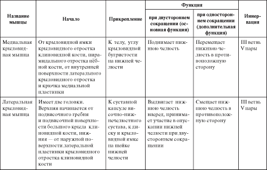
Table 10. Muscles involved in the movements of the lower jaw
Continuation of table. ten
The end of the table. ten

Typical features of the masticatory muscles
The superficial layer of the masseter muscle in brachycephaly and hameprosopic face is usually wide and low, muscle fibers diverge downward (Fig. 85); with dolichocephaly and leptoprosopic face shape, it is long and narrow, muscle fibers run parallel. The intermediate layer of this muscle with dolichocephaly and leptoprosopia protrudes more from under the posterior edge of the superficial layer than with brachycephaly and hameprosopia.
The temporalis muscle with the dolichocephalic shape of the skull is low and long, and with the brachycephalic one - high and short (see Fig. 85).
Both heads of the lateral pterygoid muscle with the brachycephalic shape of the skull are short and wide, with a narrow gap between them, with the dolichocephalic muscle - long and narrow, with a wide gap between them (Fig. 86).
The medial pterygoid muscle with the dolichocephalic shape of the skull and the leptoprosopic shape of the face is long and narrow, and with brachycephaly and chameprosopia it is low and wide (Fig. 87).
The shape of the pterygoid and masticatory muscles is determined by the shape of the ramus of the mandible and infratemporal fossa, but at the same time it corresponds to the structure of the bone components of the temporomandibular joint. This connection is especially clearly reflected in the external structure of the lateral pterygoid muscle. When the mouth is opened (lowering the lower jaw) and when the lower jaw is extended forward in people with a brachycephalic skull, the head of the joint is displaced to the apex of the flat articular tubercle, i.e. the articular path deviates slightly from the horizontal plane. This movement of the jaw head is provided by the lower head of the lateral pterygoid muscle, which lies almost horizontally. With the dolichocephalic shape of the skull, the articular head slides down the steep and high slope of the articular tubercle rather than horizontally. This movement is provided by the lower head of the lateral pterygoid muscle, the origin of which is located lower on the high lateral plate of the pterygoid process, and the muscle pulls the jaw head down rather than forward.