The anatomy of human muscles, their structure and development, perhaps, can be called the most relevant topic that arouses the maximum public interest in bodybuilding. Needless to say, it is the structure, work and function of muscles that is the topic that a personal trainer should pay special attention to. As in the presentation of other topics, we will begin the introduction to the course with a detailed study of the anatomy of muscles, their structure, classification, work and functions.
A healthy lifestyle, good nutrition and regular physical activity contribute to the development of muscles and reduce the level of body fat. The structure and work of human muscles will be understood only through a consistent study of the human skeleton first and only then the muscles. And now that we know from the article that it, among other things, performs the function of a skeleton for attaching muscles, it is high time to study what are the main muscle groups that form the human body, where they are, how they look and what functions they perform.
Above, you can see what the structure of a person's muscles looks like in the photo (3D model). Consider first the musculature of a man's body with the terms applied to bodybuilding, then the musculature of a woman's body. Looking ahead, it is worth noting that the structure of muscles in men and women has no fundamental differences, the muscles of the body are almost completely similar.
Human Muscle Anatomy
Muscles the organs of the body are called, which forms elastic tissue, and the activity of which is regulated by nerve impulses. Muscle functions include movement and movement of parts of the human body in space. Their full functioning directly affects the physiological activity of many processes in the body. Muscle work is regulated by the nervous system. It promotes their interaction with the brain and spinal cord, and also participates in the process of converting chemical energy into mechanical energy. The human body forms about 640 muscles (various methods of calculating differentiated muscle groups determine their number from 639 to 850). Below is the structure of human muscles (diagram) using the example of a male and female body.
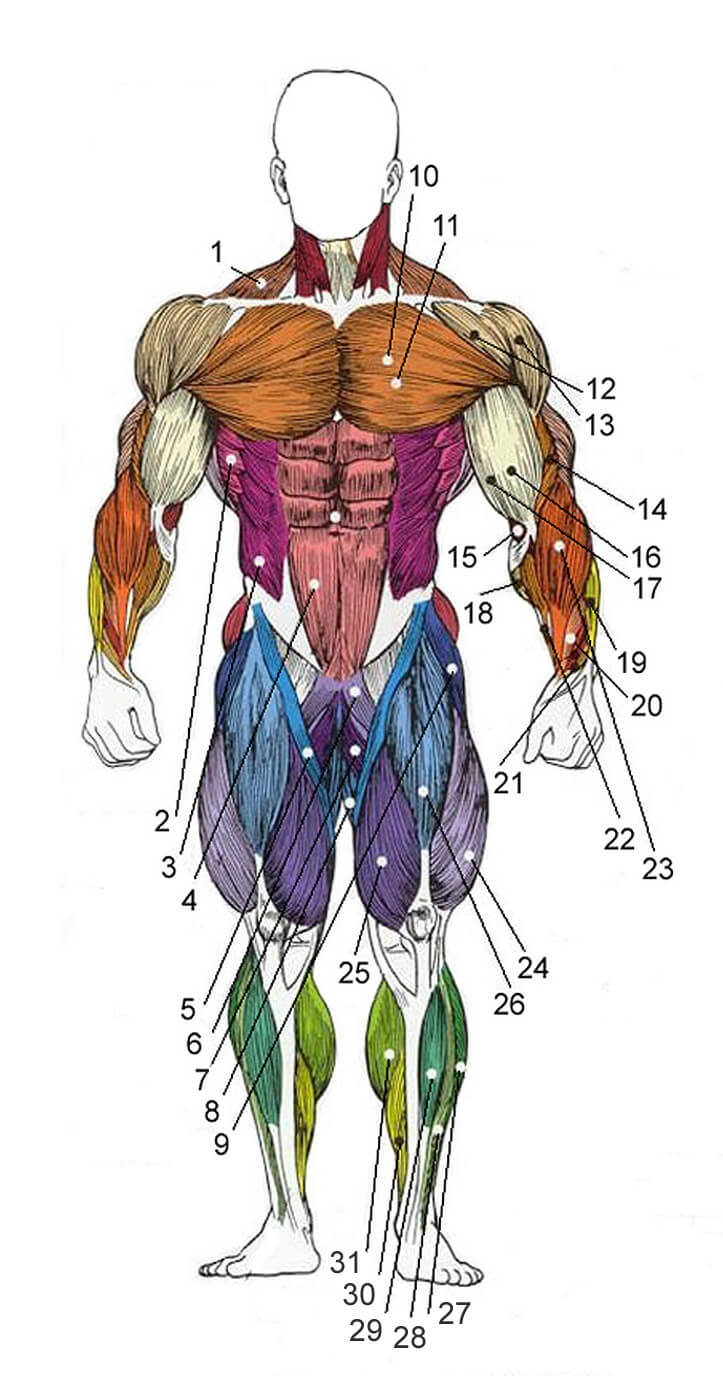
Male muscle structure, front view: 1 - trapezoid; 2 - serratus anterior muscle; 3 - external oblique muscles of the abdomen; 4 - rectus abdominis muscle; 5 - tailor muscle; 6 - comb muscle; 7 - long adductor thigh muscle; 8 - thin muscle; 9 - tensioner of the wide fascia; 10 - pectoralis major muscle; 11 - pectoralis minor; 12 - front head of the shoulder; 13 - middle head of the shoulder; 14 - brachialis; 15 - pronator; 16 - long head of the biceps; 17 - short biceps head; 18 - long palmar muscle; 19 - extensor muscle of the wrist; 20 - long adductor muscle of the wrist; 21 - long flexor; 22 - radial flexor of the wrist; 23 - brachioradial muscle; 24 - lateral thigh muscle; 25 - medial thigh muscle; 26 - rectus femoris muscle; 27 - long peroneal muscle; 28 - long extensor of the fingers; 29 - anterior tibial muscle; 30 - soleus muscle; 31 - gastrocnemius muscle
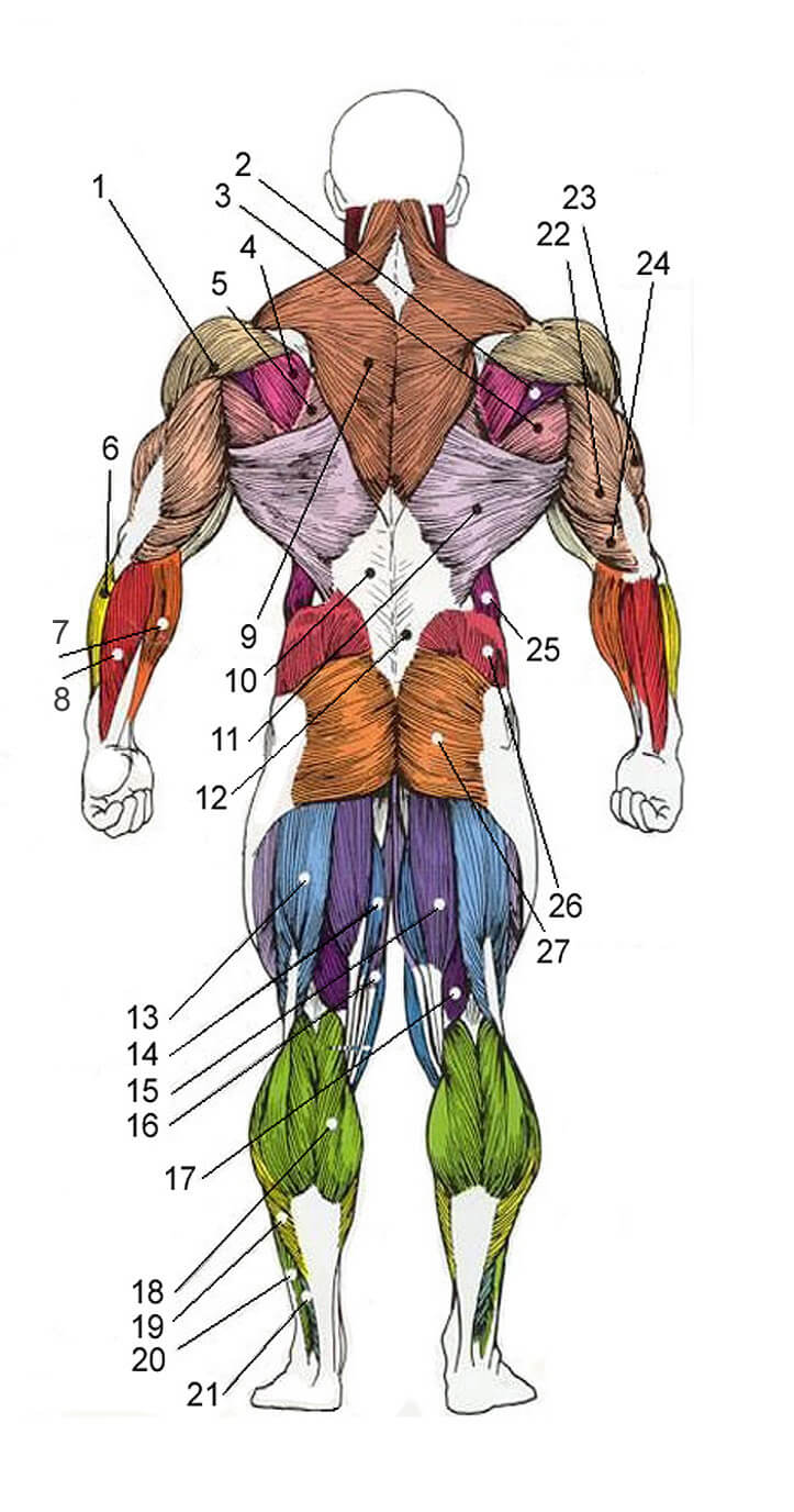
Male muscle structure, rear view: 1 - back head of the shoulder; 2 - small round muscle; 3 - large round muscle; 4 - infraspinatus muscle; 5 - rhomboid muscle; 6 - the extensor muscle of the wrist; 7 - brachioradial muscle; 8 - elbow flexor of the wrist; 9 - trapezius muscle; 10 - rectus spinous muscle; 11 - the broadest muscle; 12 - thoracolumbar fascia; 13 - thigh biceps; 14 - adductor muscle of the thigh; 15 - semitendinosus muscle; 16 - thin muscle; 17 - semi-membranous muscle; 18 - gastrocnemius muscle; 19 - soleus muscle; 20 - long peroneal muscle; 21 - muscle abducting the big toe; 22 - long triceps head; 23 - lateral head of the triceps; 24 - medial head of the triceps; 25 - external oblique muscles of the abdomen; 26 - gluteus medius muscle; 27 - gluteus maximus muscle
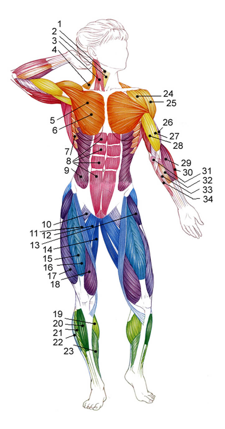
Woman's muscle structure, front view: 1 - scapular hyoid muscle; 2 - sternohyoid muscle; 3 - sternocleidomastoid muscle; 4 - trapezius muscle; 5 - pectoralis minor (not visible); 6 - pectoralis major muscle; 7 - dentate muscle; 8 - rectus abdominis muscle; 9 - external oblique muscle of the abdomen; 10 - comb muscle; 11 - tailor muscle; 12 - long adductor thigh muscle; 13 - tensioner of the wide fascia; 14 - thin muscle of the thigh; 15 - rectus femoris muscle; 16 - the intermediate broad muscle of the thigh (not visible); 17 - lateral broad muscle of the thigh; 18 - broad medial thigh muscle; 19 - gastrocnemius muscle; 20 - tibialis anterior muscle; 21 - long extensor of the toes; 22 - long tibial muscle; 23 - soleus muscle; 24 - front bundle of deltas; 25 - middle bundle of deltas; 26 - brachial muscle brachialis; 27 - a long bundle of biceps; 28 - a short bundle of biceps; 29 - brachioradial muscle; 30 - radial extensor of the wrist; 31 - round pronator; 32 - radial flexor of the wrist; 33 - long palmar muscle; 34 - elbow flexor of the wrist
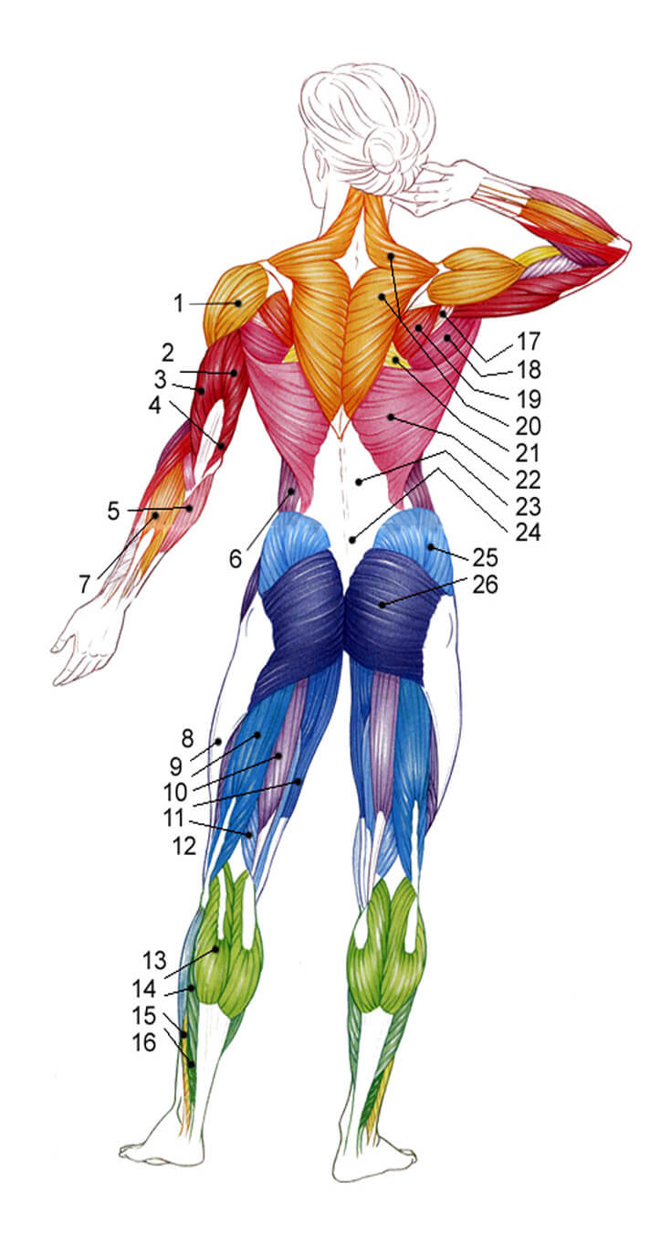
Woman's muscle structure, rear view: 1 - posterior bundle of deltas; 2 - a long bundle of triceps; 3 - lateral triceps bundle; 4 - medial triceps bundle; 5 - elbow extensor of the wrist; 6 - external oblique muscle of the abdomen; 7 - finger extensor; 8 - wide fascia; 9 - thigh biceps; 10 - semitendinosus muscle; 11 - thin muscle of the thigh; 12 - semi-membranous muscle; 13 - gastrocnemius muscle; 14 - soleus muscle; 15 - short peroneal muscle; 16 - long flexor of the thumb; 17 - small round muscle; 18 - large round muscle; 19 - infraspinatus muscle; 20 - trapezius muscle; 21 - rhomboid muscle; 22 - the broadest muscle; 23 - extensors of the spine; 24 - thoracolumbar fascia; 25 - gluteus maximus muscle; 26 - gluteus maximus muscle
Muscles are quite varied in shape. Muscles that share a common tendon but have two or more heads are called biceps (biceps), triceps (triceps), or quadriceps (quadriceps). The functions of the muscles are also quite diverse, these are flexors, extensors, abductors, adductors, rotators (inward and outward), lifting, lowering, straightening and others.
Types of muscle tissue
The characteristic features of the structure make it possible to classify human muscles into three types: skeletal, smooth and cardiac.
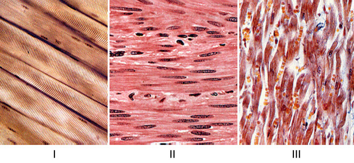
Types of human muscle tissue: I - skeletal muscles; II - smooth muscles; III- cardiac muscle
- Skeletal muscle. The contraction of this type of muscle is completely controlled by the person. Combined with the human skeleton, they form the musculoskeletal system. This type of muscle is called skeletal precisely because of their attachment to the bones of the skeleton.
- Smooth muscles. This type of tissue is found in the cells of internal organs, skin and blood vessels. The structure of human smooth muscles implies their finding mostly in the walls of hollow internal organs, such as the esophagus or bladder. They also play an important role in processes not controlled by our minds, such as intestinal motility.
- Heart muscle (myocardium). The autonomic nervous system controls the work of this muscle. Its contractions are not controlled by human consciousness.
Since the contraction of smooth and cardiac muscle tissue is not controlled by human consciousness, the emphasis in this article will focus on skeletal muscles and their detailed description.
Muscle structure
Muscle fiber is a structural element of muscles. Individually, each of them is not only a cellular, but also a physiological unit that is capable of contracting. The muscle fiber has the appearance of a multinucleated cell, the fiber diameter is in the range from 10 to 100 microns. This multinucleated cell is located in a membrane called the sarcolemma, which in turn is filled with sarcoplasm, and myofibrils are already in the sarcoplasm.
Myofibril is a filamentous formation that consists of sarcomeres. The thickness of myofibrils is usually less than 1 micron. Taking into account the number of myofibrils, white (they are also fast) and red (they are also slow) muscle fibers are usually distinguished. White fibers contain more myofibrils, but less sarcoplasm. It is for this reason that they shrink faster. Red fibers contain a lot of myoglobin, which is why they got this name.
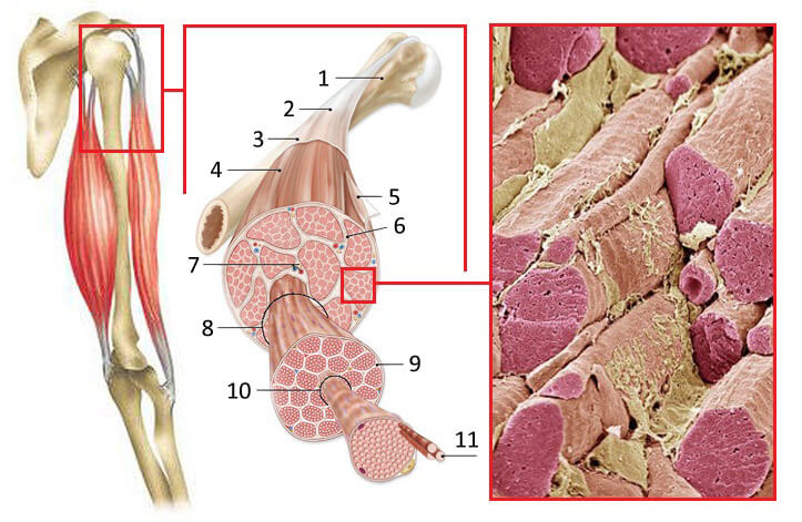
Internal structure of a human muscle: 1 - bone; 2 - tendon; 3 - muscle fascia; 4 - skeletal muscle; 5 - fibrous membrane of skeletal muscle; 6 - connective tissue membrane; 7 - arteries, veins, nerves; 8 - beam; 9 - connective tissue; 10 - muscle fiber; 11 - myofibril
Muscle work is characterized by the fact that the ability to contract faster and stronger is characteristic of white fibers. They can develop force and the speed of contraction is 3-5 times higher than slow fibers. Physical activity of the anaerobic type (work with weights) is performed mainly by fast muscle fibers. Long-term aerobic physical activity (running, swimming, cycling) is performed mainly by slow muscle fibers.
Slow fibers are more resistant to fatigue, while fast fibers are not adapted to prolonged physical activity. As for the ratio of fast and slow muscle fibers in human muscles, their number is approximately the same. In most of both sexes, about 45-50% of the muscles of the limbs are slow muscle fibers. There are no significant sex differences in the ratio of different types of muscle fibers in men and women. Their ratio is formed at the beginning of a person's life cycle, in other words, it is genetically programmed and practically does not change until old age.
Sarcomeres (constituents of myofibrils) are formed by thick myosin filaments and thin actin filaments. Let's dwell on them in more detail.
Actin- a protein that is a structural element of the cytoskeleton of cells and has the ability to contract. It consists of 375 amino acid residues and makes up about 15% of muscle protein.
Myosin- the main component of myofibrils - muscle contractile fibers, where its content can be about 65%. The molecules are formed by two polypeptide chains, each of which contains about 2000 amino acids. Each of these chains has a so-called head at the end, which includes two small chains of 150-190 amino acids.
Actomyosin- a complex of proteins formed from actin and myosin.
FACT. For the most part, muscles are made up of water, proteins and other components: glycogen, lipids, nitrogen-containing substances, salts, etc. The water content ranges from 72-80% of the total muscle mass. Skeletal muscle consists of a large number of fibers, and what is characteristic, the more there are, the stronger the muscle.
Muscle classification
The human muscular system is characterized by a variety of muscle shapes, which in turn are divided into simple and complex. Simple: fusiform, straight, long, short, wide. The multi-headed muscles can be classified as complex. As we have already said, if the muscles have a common tendon, and there are two or more heads, then they are called two-headed (biceps), triceps (triceps) or quadriceps (quadriceps), as well as multi-tendon and digastric muscles. The following types of muscles with a certain geometric shape are also complex: square, deltoid, soleus, pyramidal, round, dentate, triangular, rhomboid, soleus.
Main functions muscles are flexion, extension, abduction, adduction, supination, pronation, lifting, lowering, straightening and more. The term supination means outward rotation, and the term pronation means inward rotation.
In the direction of the fibers muscles are divided into: straight, transverse, circular, oblique, single-pinnate, two-pinnate, multi-pinnate, semitendinosus and semimembranous.
In relation to the joints, taking into account the number of joints through which they are thrown: single-joint, double-joint and multi-joint.
Muscle work
In the process of contraction, the actin filaments penetrate deeply into the gaps between the myosin filaments, and the length of both structures does not change, but only the total length of the actomyosin complex is reduced - this method of muscle contraction is called sliding. The sliding of actin filaments along myosin filaments requires energy, and the energy required for muscle contraction is released as a result of the interaction of actomyosin with ATP (adenosine triphosphate). In addition to ATP, water plays an important role in muscle contraction, as well as calcium and magnesium ions.
As already mentioned, muscle work is completely controlled by the nervous system. This suggests that their work (contraction and relaxation) can be controlled consciously. For the normal and full functioning of the body and its movement in space, muscles work in groups. Most of the muscle groups of the human body work in pairs, and perform opposite functions. It looks in such a way that when the "agonist" muscle contracts, the "antagonist" muscle is stretched. The same is true vice versa.
- Agonist- a muscle that performs a specific movement.
- Antagonist- a muscle performing the opposite movement.
Muscles have the following properties: elasticity, stretching, contraction. Elasticity and stretching give the muscles the opportunity to change in size and return to their original state, the third quality makes it possible to create force at its ends and lead to shortening.
Nerve stimulation can cause the following types of muscle contraction: concentric, eccentric and isometric. Concentric contraction occurs in the process of overcoming the load when performing a given movement (lifting up when pulling up on the bar). Eccentric contraction occurs in the process of slowing down the movements in the joints (lowering down when pulling up on the bar). Isometric contraction occurs at the moment when the force created by the muscles is equal to the load exerted on them (keeping the body hanging on the bar).
Muscle functions
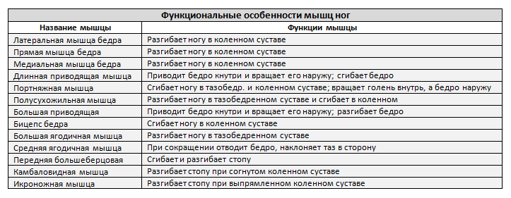
Knowing the name and where this or that muscle or muscle group is located, we can proceed to the study of the block - the function of human muscles. Below in the table we will look at the most basic muscles that are trained in the gym. Typically, six main muscle groups are trained: chest, back, legs, shoulders, arms and abs.
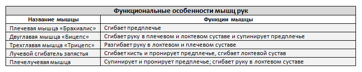
FACT. The largest and strongest muscle group in the human body is the legs. The largest muscle is the gluteus maximus. The strongest is the calf, it can hold up to 150 kg.
Conclusion
In this article, we examined such a complex and voluminous topic as the structure and function of human muscles. Speaking of muscles, we of course also mean muscle fibers, and the involvement of muscle fibers in the work involves the interaction of the nervous system with them, since the execution of muscle activity is preceded by the innervation of motor neurons. It is for this reason that in our next article we will move on to examining the structure and functions of the nervous system.